
Product data from the official website biossci.com · For selection reference only; specs & stock subject to the vendor.
Quick Specs
BBL-0104
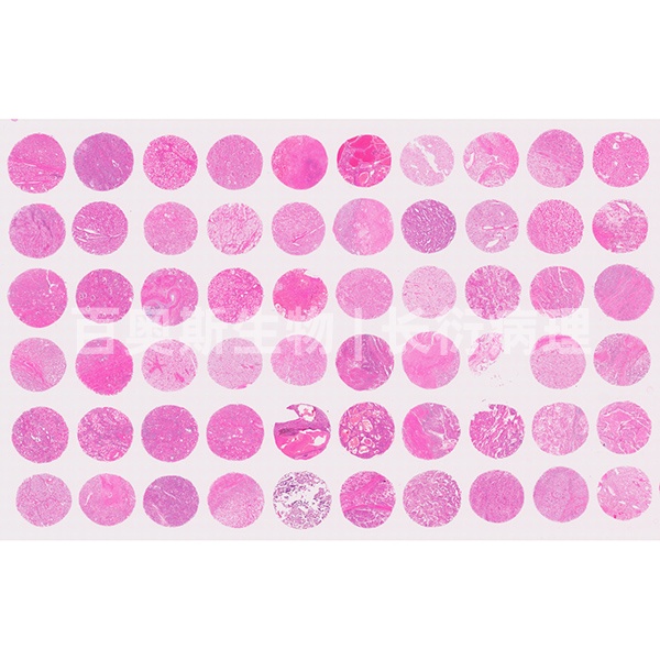
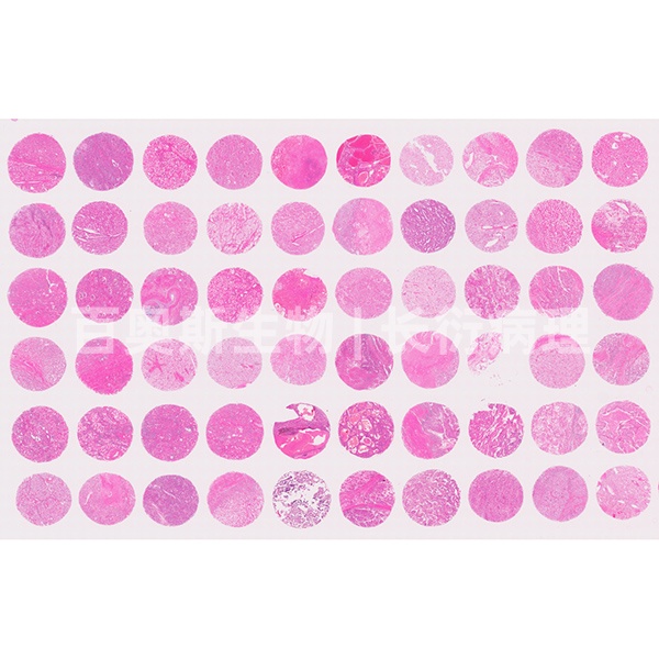
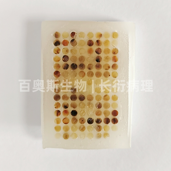
H&E staining of tissue microarray
| Spec | — |
|---|---|
| Fixation | — |
| Applications | Zhang |
| Price | ¥20.00 |
| Stock | Please consult |
| Lead Time | 7天内 |
👁 Views 46 · 组织芯片制作 · 2026-10-05
Product Specifications
| Cat. No. | BBL-0104 |
|---|---|
| Cases | — |
| Cores | — |
| Core diameter | — |
| Section thickness | — |
| Fixation | — |
| Storage | — |
| Applications | Zhang |
Product Details
Service Introduction:
Hematoxylin-eosin staining (hematoxylin-eosin staining), abbreviated as HE staining, is currently the most widely used histological staining method because it is relatively simple and can clearly reveal many different tissue structures. Hematoxylin staining solution is basic and mainly stains chromatin in the nucleus and ribosomes in the cytoplasm purplish blue; eosin is an acidic dye that mainly stains components in the cytoplasm and extracellular matrix red. HE staining is the most basic and most widely used technique in teaching and research in histology, embryology, and pathology. This project provides HE staining for tissue microarrays.
Main Applications:
It is mainly used for staining various cells, paraffin-embedded tissue sections, frozen sections, smears, and imprint preparations.
Test Principle:
Hematoxylin-eosin staining solution is mainly used to reveal the general morphological structure of normal components and lesions in various tissues for comprehensive observation. The nucleus in cells is composed of acidic substances and has a strong affinity for basic dyes (hematoxylin), whereas the cytoplasm is the opposite: it contains basic substances and has a greater affinity for acidic dyes (eosin). Therefore, after cells or tissue sections are stained with hematoxylin-eosin staining solution, nuclei are stained bright blue-purple by hematoxylin, while cytoplasm, muscle fibers, collagen fibers, etc. are stained varying degrees of red, and erythrocytes appear orange-red.
Experimental Procedure:
Deparaffinization to water - hematoxylin staining - eosin staining - dehydration and mounting - microscopic examination.
Sample Requirements and Transport Conditions:
1. Provide properly fixed and qualified samples. After tissue removal, immediately fix with an appropriate fixative; the volume of fixative is recommended to be more than 10–15 times the tissue volume. Fresh specimens should be fixed in appropriate containers for 24–48 h; large specimens should be fixed for 12 h before being cut and fixed further. Specimens should be fixed at room temperature (around 24°C) or refrigerated (4°C); do not freeze. Fixed samples should be transported at room temperature for submission.
2. Smears and cell coverslips must be fully fixed, and paraffin sections must be fully deparaffinized before the experiment.
Inquiry · BBL-0104
🎓 相关高校、实验室、医院与科研需求
Contact the Webmaster
WeChat ID
Open WeChat → Contacts → Add Contact → paste the ID above.