
Product data from the official website biossci.com · For selection reference only; specs & stock subject to the vendor.
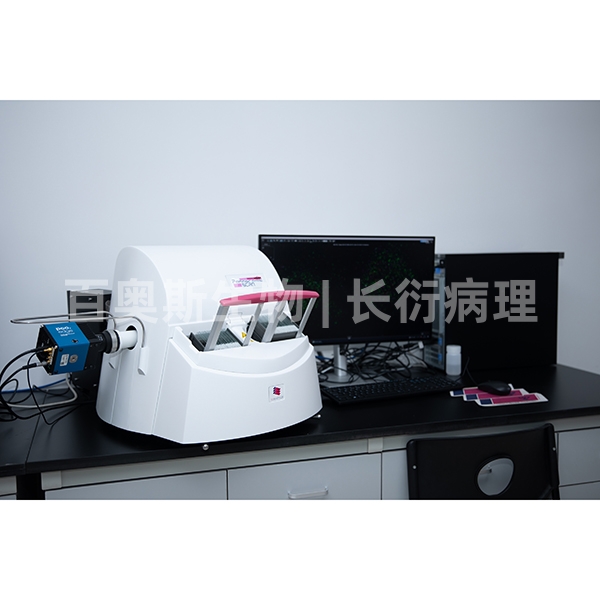
Quick Specs
BBL-0109
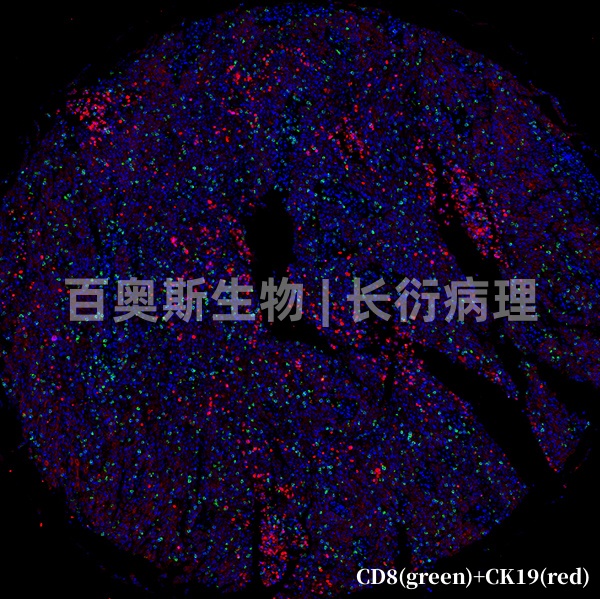
Tissue microarray triple immunofluorescence (IF) staining
| Spec | — |
|---|---|
| Fixation | — |
| Applications | Zhang |
| Price | ¥1,200.00 |
| Stock | Please consult |
| Lead Time | 7天内 |
👁 Views 72 · 组织芯片免疫荧光 · 2026-10-05
Product Specifications
| Cat. No. | BBL-0109 |
|---|---|
| Cases | — |
| Cores | — |
| Core diameter | — |
| Section thickness | — |
| Fixation | — |
| Storage | — |
| Applications | Zhang |
Product Details
Service introduction:
Immunofluorescence technique is a technique established on the basis of immunology, biochemistry, and microscopy. The method of using fluorescent antibodies to trace or examine corresponding antigens is called the fluorescent antibody method; the method of using known fluorescent antigen markers to trace or examine corresponding antibodies is called the fluorescent antigen method. These two methods are collectively referred to as immunofluorescence techniques. However, in practical work, the fluorescent antigen technique is rarely used, so the fluorescent antibody technique is often referred to as immunofluorescence technique.
Labeling three antigens on a single section is referred to as immunofluorescence (IF) triple labeling. Conventional immunofluorescence triple labeling requires that primary antibodies be derived from different sources; if they are from the same source, secondary antibodies cannot distinguish them. Different primary antibodies are correspondingly labeled with different colors and can be used to localize three different antigens. Tissue microarray immunofluorescence can arrange dozens to hundreds of small tissues neatly on a single slide for staining, keeping the experimental conditions of tissue samples on the microarray consistent, saving time and reagents, and facilitating unified analysis after scanning. Compared with single-section staining, both time and cost can be reduced. The disadvantage is that the selected tissue area is relatively small and cannot comprehensively reflect the expression status of individual samples. The tissue microarray immunofluorescence triple labeling procedure is basically the same as the conventional tissue immunofluorescence triple labeling procedure, but because the samples are relatively precious, the operational procedures are more stringent.
Main applications:
Immunofluorescence staining and imaging analysis are indispensable detection techniques for studying tissue morphology and in situ antigen expression in tissues and are widely used in various fields of clinical pathology and medical and biological research. Any substance that can serve as an antigen or hapten, such as proteins, polypeptides, amino acids, polysaccharides, phospholipids, enzymes, hormones, pathogens, and receptors, can be detected at the cellular and subcellular levels.
Detection principle:
Immunofluorescence technique is based on the principle of specific binding between antigens and antibodies. First, known antibodies (or antigens) are labeled with fluorophores, and then these fluorescent antibodies (or antigens) are used as probes to detect corresponding antigens (or antibodies) in fixed cells or tissues. Under a fluorescence microscope, when the fluorophore in the antigen-antibody complex is irradiated by excitation light, it emits fluorescence of a certain wavelength, thereby determining the nature and localization of the target antigen (or antibody). Quantitative techniques such as flow cytometry can also be used to measure its content.
Experimental procedure:
Dewaxing and rehydration - antigen retrieval - serum blocking - primary antibody incubation - secondary antibody incubation - DAPI nuclear staining - mounting - microscopy.
Sample requirements and transport conditions:
1. Provide properly fixed and qualified samples. After tissue removal, immediately fix the tissue in an appropriate fixative. The volume of fixative should be greater than 10-15 times the tissue volume. Fresh specimens should be fixed in appropriate containers for 24-48 h; large specimens should be fixed for 12 h, then cut open and fixed. Specimens should be fixed at room temperature (about 24°C) or refrigerated (4°C); do not freeze. Fixed samples should be transported at room temperature.
2. Smears and coverslip cell preparations must be fully fixed; paraffin sections must be fully dewaxed before the experiment.
3. Provide accurate antibody information.
Precautions:
Inquiry · BBL-0109
Related Products
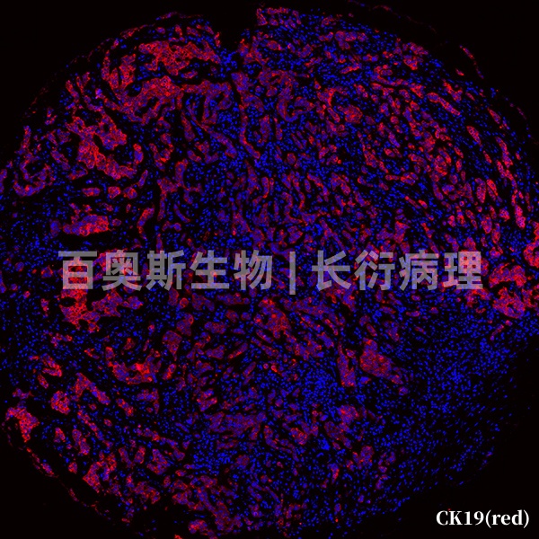
BBL-0107
Tissue microarray immunofluorescence (IF) single labeling
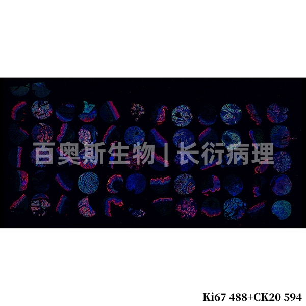
BBL-0108
Tissue microarray immunofluorescence (IF) double labeling
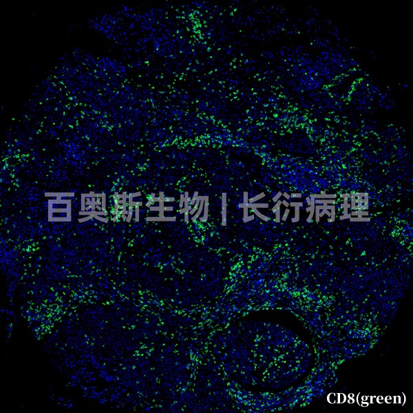
BBL-0110
Tissue microarray immunofluorescence single labeling (TSA method)
BBL-0111
Tissue microarray immunofluorescence double-labeling (TSA method)
🎓 相关高校、实验室、医院与科研需求
Contact the Webmaster
WeChat ID
Open WeChat → Contacts → Add Contact → paste the ID above.