
Product data from the official website biossci.com · For selection reference only; specs & stock subject to the vendor.
Quick Specs
BBL-0110
Tissue microarray immunofluorescence single labeling (TSA method)
| Spec | — |
|---|---|
| Fixation | — |
| Applications | Zhang |
| Price | ¥500.00 |
| Stock | Please consult |
| Lead Time | 7天内 |
👁 Views 80 · 组织芯片免疫荧光 · 2026-10-05
Product Specifications
| Cat. No. | BBL-0110 |
|---|---|
| Cases | — |
| Cores | — |
| Core diameter | — |
| Section thickness | — |
| Fixation | — |
| Storage | — |
| Applications | Zhang |
Product Details
Service Introduction:
Multiplex immunohistochemistry (mIHC) is an important technical method that has emerged in the field of pathology research in recent years. It is a multiplex sequential immunostaining technique based on tyramide signal amplification (TSA), which can detect multiple target molecules in situ in cell or tissue samples. By studying the combinations and spatial relationships of these targets, their interaction mechanisms can be elucidated. This technology can not only greatly improve the sensitivity of detected signals, but also solve the challenge of detecting multiple biomarkers from the same species on a single section. Through quantitative pathological analysis, information such as the types, components, and expression levels of various targets in situ in tissue cells can be obtained, as well as the spatial localization, qualitative and quantitative information of each target and its interactions. Comprehensive, systematic, intuitive, and scientific interpretation of this information is of great significance for studying the mechanisms of disease occurrence and progression, and for exploring effective methods for disease diagnosis and treatment.
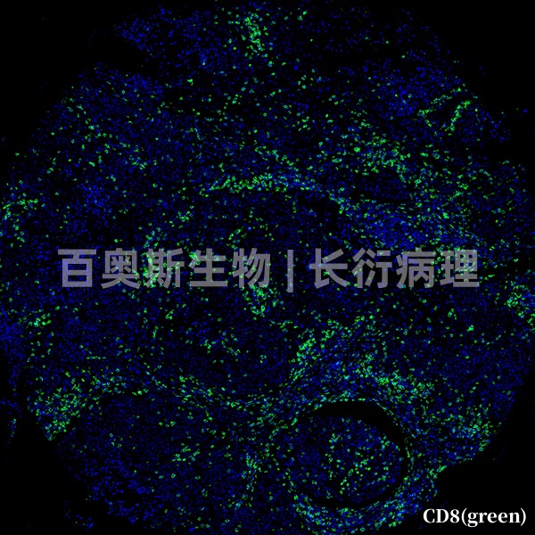
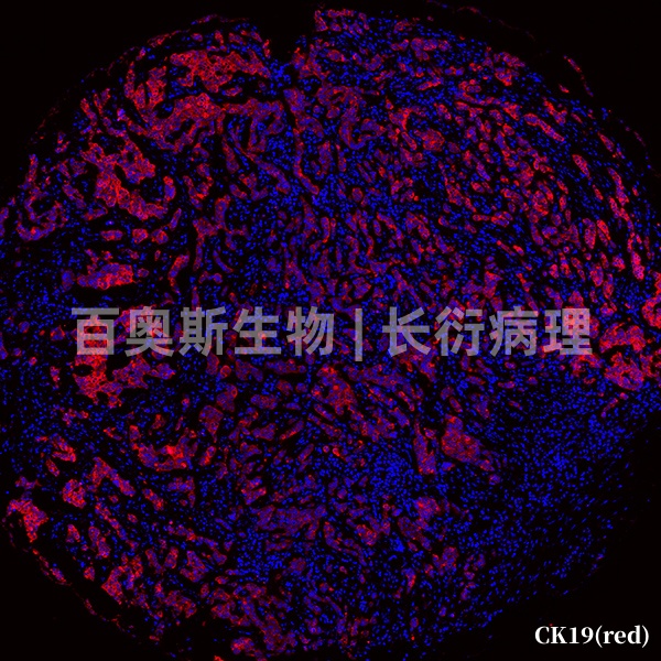
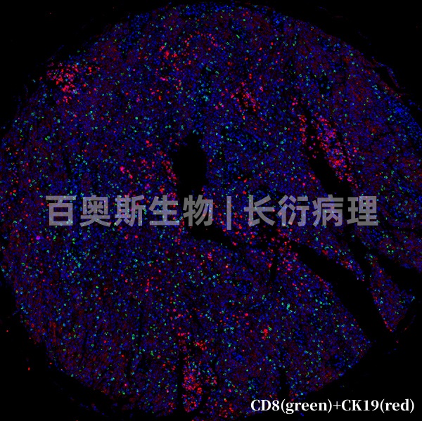
In this project, a single antigen is labeled on one chip by the TSA method, usually with red or green fluorescence, and nuclei are labeled with DAPI.
Main Applications:
1. Dual advantages of in situ visualization and quantitative analysis.
2. More comprehensive analysis: simultaneous analysis of drug targets and the tumor microenvironment.
3. Free combination of different targets, multi-target co-localization staining, and signal separation.
4. Display spatial positional relationships and distinguish different cell subpopulations.
Detection Principle:
In the presence of hydrogen peroxide, enzymes such as horseradish peroxidase and alkaline phosphatase can catalyze a tyramide substrate into a highly reactive, short-lived intermediate. Within a short period, the intermediate can covalently bind to electron-rich surfaces of surrounding proteins to form tyramide complexes. Because adjacent proteins, such as HRP, antibodies, and target antigens, contain abundant tyrosine residues, large amounts of signal are enriched and the signal is effectively amplified.
Experimental Workflow:
Deparaffinization and rehydration - antigen retrieval - inactivation of endogenous peroxidase - serum blocking - primary antibody incubation - secondary antibody incubation - fluorophore labeling - elution - DAPI nuclear staining - mounting - microscopic examination.
Sample Requirements and Transport Conditions:
1. Provide properly fixed and qualified samples. After tissue excision, immediately fix the tissue in an appropriate fixative; the volume of fixative is recommended to be at least 10–15 times the tissue volume. Fresh specimens should be fixed in appropriate containers for 24–48 h; large specimens should be fixed for 12 h and then cut open for further fixation. Samples should be fixed at room temperature (around 24°C) or refrigerated (4°C); do not freeze. Fixed samples should be transported at room temperature.
Inquiry · BBL-0110
Related Products
BBL-0107
Tissue microarray immunofluorescence (IF) single labeling
BBL-0108
Tissue microarray immunofluorescence (IF) double labeling
BBL-0109
Tissue microarray triple immunofluorescence (IF) staining
BBL-0111
Tissue microarray immunofluorescence double-labeling (TSA method)
🎓 相关高校、实验室、医院与科研需求
Contact the Webmaster
WeChat ID
Open WeChat → Contacts → Add Contact → paste the ID above.